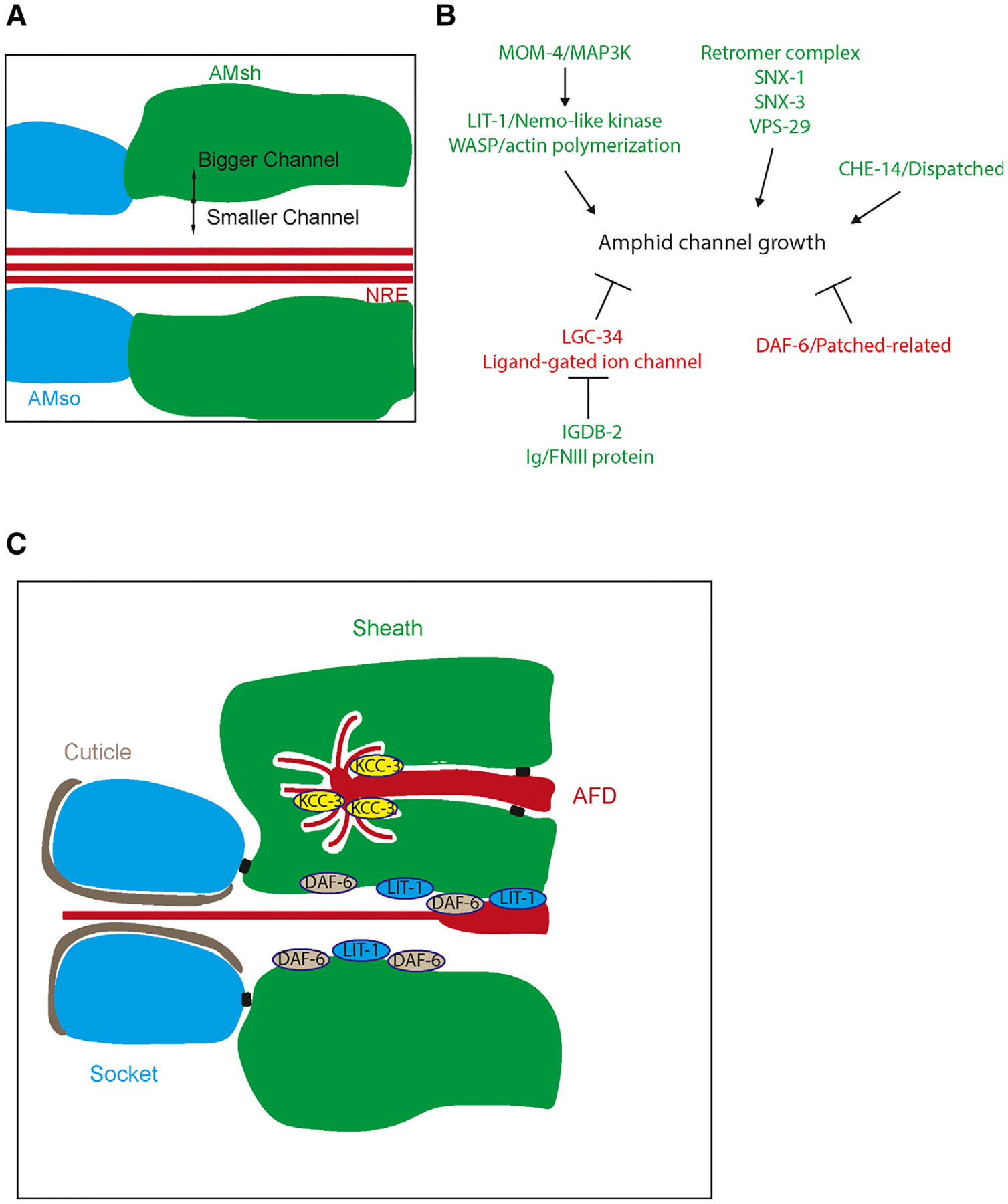
Fig. 2.

(A) Schematic of the amphid channel. AMsh (green) and AMso (blue) form a channel that certain neuron receptive endings (NREs) sit in. Regulation of AMsh growth in this area can determine the size of the lumen. (B) Model for amphid channel morphogenesis. Genes that increase channel size are colored green while those that decrease channel size are colored red. (C) Distinct subcellular domains are generated by the AMsh depending on its specific neuronal partners. For example, KCC-3 is localized to the AMsh pocket interacting with the AFD neuron while DAF-6 and LIT-1 are localized to the main amphid channel.
