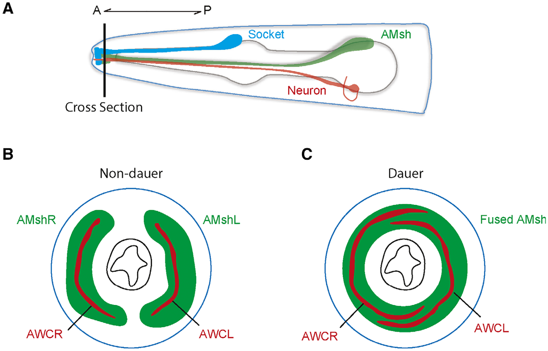
Fig. 4.

(A) Schematic of the head of the worm showing the AMsh glia. Horizontal line indicates cross section visualized in (B) and (C). (B-C) Sections showing the two bilateral AMsh glia (green) and AWC neurons (red) in non-dauer (B) and dauer (C) conditions. In dauer animals, the two AMsh glia fuse while the AMC receptive endings expand.
