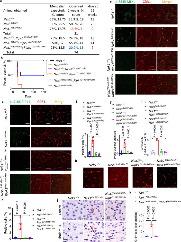
Fig. 1. Rescue of BBB damage in Nek1Kat2J mice by RIPK1 inhibition.
a, b The surviving progenies (at 2 weeks and 22 weeks of age) from the following matings: 8 female Nek1Kat2J/+ mice were mated with 4 male Nek1Kat2J/+ mice; 8 female Nek1Kat2J/+; Ripk1D138N/D138N mice were mated with 4 male Nek1Kat2J/+; Ripk1D138N/D138N mice. The % of survival was ploted (b). c–f, Cerebral cortical sections from the mice (3 weeks old, female) were immunostained with anti-p-S166 RIPK1 (c) and anti-p-S345 MLKL (e). The fluorescent signals of p-S166 RIPK1 (d) and p-S345 MLKL (f) were quantified. Mean ± SD. n = 3 sections per group for p-S166 RIPK1. n = 6 sections per group for p-S345 MLKL. Bar = 50 µm. One-way ANOVA with Dunnett’s test. g Mice (3 weeks old) were inraperitoneally injected with 2% (w/v) Evans blue solution (70 mg/kg) and followed by PBS perfusion. Extravasated Evans blue levels in total brain lysates were measured by a plate reader. Mean ± SD. n = 3 mice per group. One-way ANOVA with Dunnett’s test. h, i Mice (3 weeks old) were intravenously injected with 10kD-tetramethyrhodamine (TMR)-labeled Dextran and then sacrificed. The cryo-sections of cerebral cortex were imaged by confocal microscope. Bar = 20 µm. Fluorescent intensity of diffused dextran around cerebral vessels was quantified using ImageJ. The fluorescent intensity of all groups was nomalized to Nek1+/+ mice (mean ± SD, n = 4 sections per group) and graphed in (i). One-way ANOVA with Dunnett’s test. j, k Representative images of Gr1 immunohistostaining (brown) of cerebral cortex and thalamus sections (3 weeks old mice). Bar = 20 µm.Number of Gr1 positive cells per sections were presented as mean± SD. n = 3 sections per group. One-way ANOVA with Dunnett’s test.

