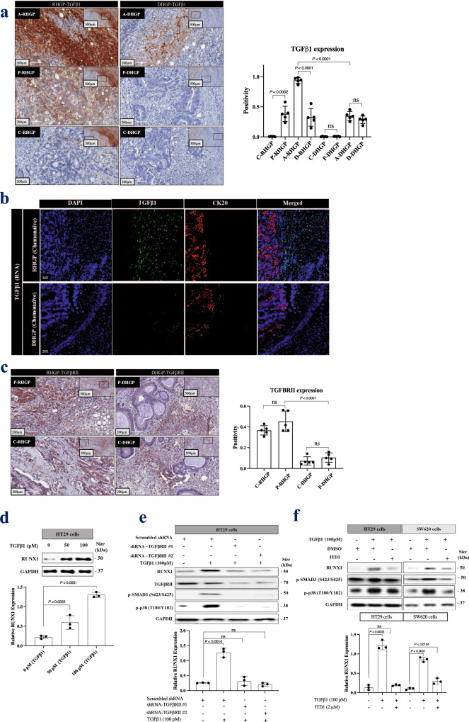
Fig. 2. TGFβ1 regulates RUNX1 expression in cancer cells through TGFβRII.
a Left panel represents immunohistochemical staining of RHGP (n = 5) and DHGP (n = 5) chemonaïve CRCLM specimens using TGFβ1 antibody. The right panel shows the positivity [total number of positive pixels/total number of pixels] was measured using an optimized Aperio algorithm (mean + SD). b. Fluorescence in situ hybridization (FISH) for TGFβ1 mRNA (green) expression in chemonaïve CRCLM lesions overlapped with CK20 (cytokeratin 20, red) antibody. c The left panel shows immunohistochemistry staining of chemonaïve CRCLM lesions with TGFβRII antibody. The right panel shows the positivity [total number of positive pixels/total number of pixels] was measured using an optimized Aperio algorithm (mean + SD). d Western blot of RUNX1 in HT29 cancer cells upon treatment with recombinant TGFβ1 for 24 h (top panel). e Western blot of RUNX1 and TGFβRII in HT29 cancer cells expressing either scrambles shRNA or shRNA against TGFβRII in the presence or absence of recombinant TGFβ1 for 24 h (top panel). f Western blot of RUNX1 in HT29 and SW620 cancer cells in the presence or absence of recombinant TGFβ1 individually or TGFβ1 with 2 μM of TGFβRII inhibitor (ITD1) for 24 h (top panel). The bottom panels represent the intensity of the bands (n = 3). C-RHGP Central tumor cells in RHGP lesions, P-RHGP Peripheral tumor cells in RHGP lesions, A-RHGP Adjacent hepatocytes to tumor lesion in RHGP, D-RHGP Distal hepatocytes to tumor lesion in RHGP, C-DHGP Central tumor cells in DHGP, P-DHGP Peripheral tumor cells in DHGP lesions, A-DHGP Adjacent hepatocytes to tumor lesion in DHGP, D-DHGP Distal hepatocytes to tumor lesion in DHGP. Data are presented as the mean ± SD.

