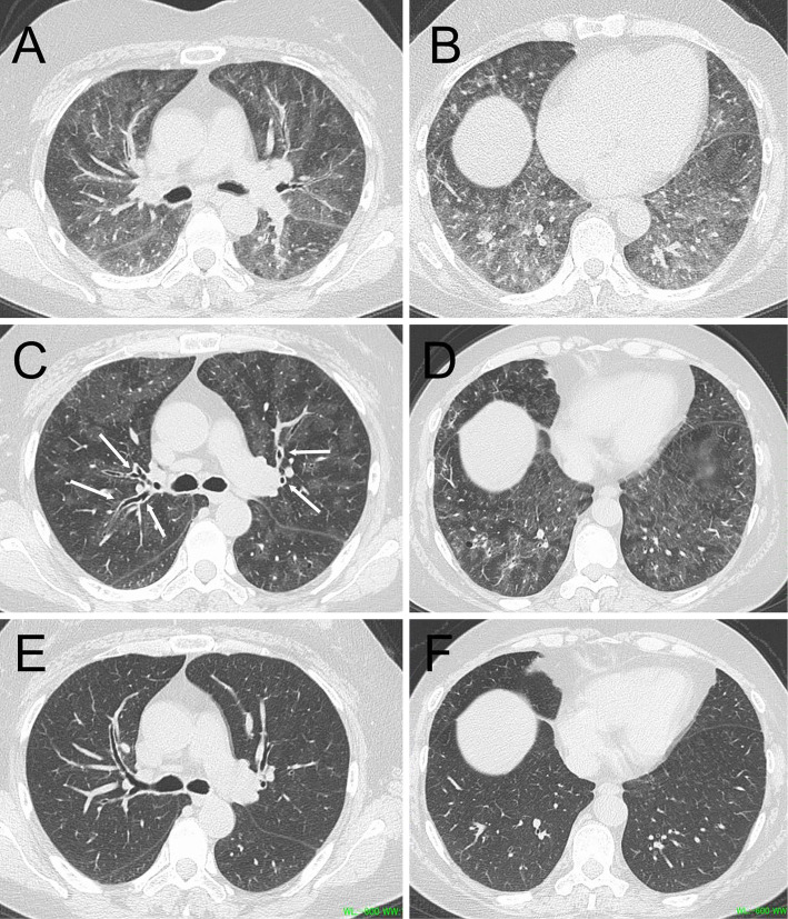
Figure 1.
Chest high-resolution computed tomography (HRCT) images. Expiratory CT on admission showed an extensive mosaic attenuation pattern in both the upper (A) and lower lobes (B). Inspiratory CT on admission revealed numerous ground-glass opacities predominantly distributed around the bronchovascular bundles in both the upper (C) and lower lobes (D), along with peribronchial cuffing (arrows). Ground-glass opacities had disappeared on day 51 (E, F).

