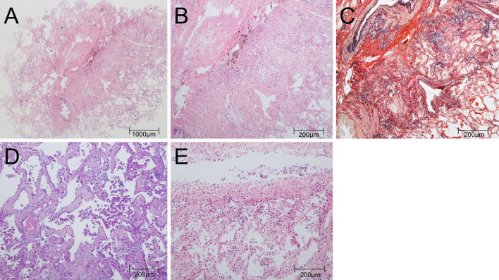
Figure 2.
Histological images. (A) The lesion was characterized by diffuse thickening of the alveolar septum and moderate intraluminal organization (right B8a) [Hematoxylin and Eosin (H&E) staining, ×4]. (B) Mild infiltration of lymphocytes and eosinophils was observed (right B8a) (H&E staining, ×20). (C) In the alveolar septum, collagen deposition and the growth of elastic fibers were observed (right B8a) (Elastica van Gieson stain, ×20). (D) In some alveolar cavities, a cluster of eosinophilic macrophages was observed, but no apparent granulomas were seen (right B8a) (periodic acid-Schiff stain, ×20). (E) Mild infiltration of lymphocytes and plasma cells into the bronchial walls was also observed (right B9a) (H&E staining, ×20).

