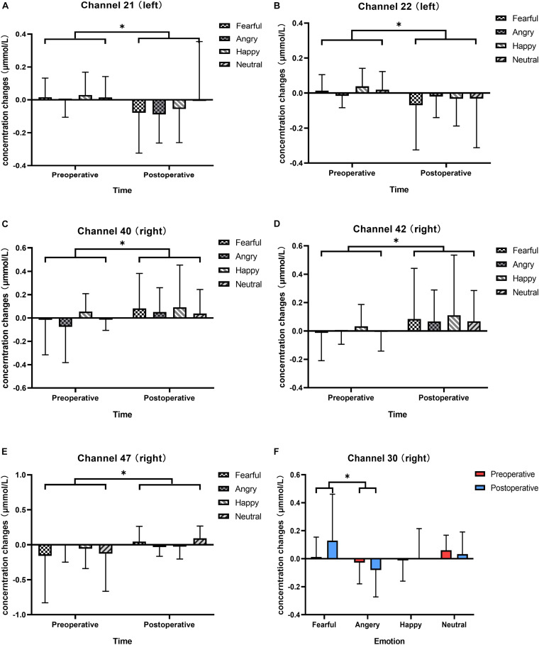
FIGURE 4.
Changes in the responses of cortical oxyhemoglobin to four vocal emotional stimuli (the white grid pattern represents fearful emotion, the gray grid pattern represents angry emotion, the white diagonal pattern represents happy emotion, and the gray diagonal pattern represents neutral emotion) in the two-period test (red represents the preoperative test, blue represents the postoperative test). The six subplots show channels 21, 22 (located in the left pre-motor and supplementary motor cortex), 30 (located in the right supramarginal gyrus of Wernicke’s area), 40, 42 (located in the middle temporal gyrus), and 47 (located in the superior temporal gyrus). (A,B) In channels 21 and 22 (located in the left pre-motor and supplementary motor cortex), there is a significant difference in cortical activation elicited by vocal emotional stimulation in the preoperative and postoperative tests. (C,D) In channels 40 and 42 (located in the right middle temporal gyrus), there is a significant difference in cortical activation elicited by vocal emotional stimulation in the preoperative and postoperative tests. (E) In channel 47 (located in the right superior temporal gyrus), there is a significant difference in cortical activation elicited by vocal emotional stimulation in the preoperative and postoperative tests. (F) In channel 30 (located in the right supramarginal gyrus of Wernicke’ s area), cortical activation elicited by fear is significantly different than that elicited by anger. ∗p < 0.05.

