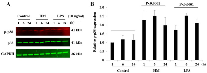
Figure 5.
Effect of HM on the phosphorylation of p38 MAPK. Representative western blotting. (A) showing the effect of HM and LPS (10 µg/ml. on p38 MAPK phosphorylation in THP-1 cells after 1-, 6- and 24-h stimulation compared with the untreated cells. The bar graph. (B) shows the relative protein expression levels of p-p38 calculated as a ratio of total p38 expression (the loading control). The results are presented as the mean ± SD of three independent experiments. P-values signify a statistically significant difference compared with the control group. HM, herbal melanin; LPS, lipopolysaccharide; IL-1β, interleukin-1β; p-, phosphorylated.

