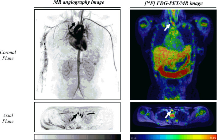
Figure 2.
Illustrative case of a 17-year-old female patient with childhood-onset Takayasu Arteritis. Left: Angioresonance of aorta and its branches after intravenous administration of gadolinium. Arrows indicates an occlusion at the third portion of the brachiocephalic trunk. Right: Arterial inflammation grade III as assessed by 18F-fluorodeoxyglucose-positron emission tomography/magnetic resonance image at the brachiocephalic trunk (SUVmax=2.38).

