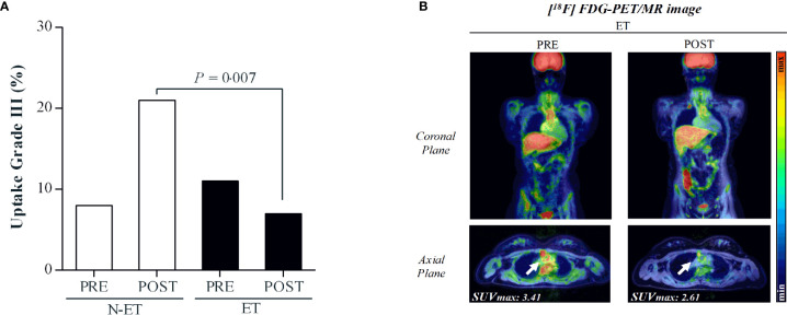
Figure 3.
Panel (A): Distribution of vessel segments in grade III, representing severe inflammation. P=0.007 for between-group comparison at POST. Panel (B): Illustrative case of a 20-year-old patient with childhood-onset Takayasu Arteritis. The [18F] FDG PET/MR imaging revealed grade III inflammation in the ascending aorta artery before the intervention (SUVmax=3.41). Following the exercise training program, the uptake decreased to grade II (SUVmax=2.61).

