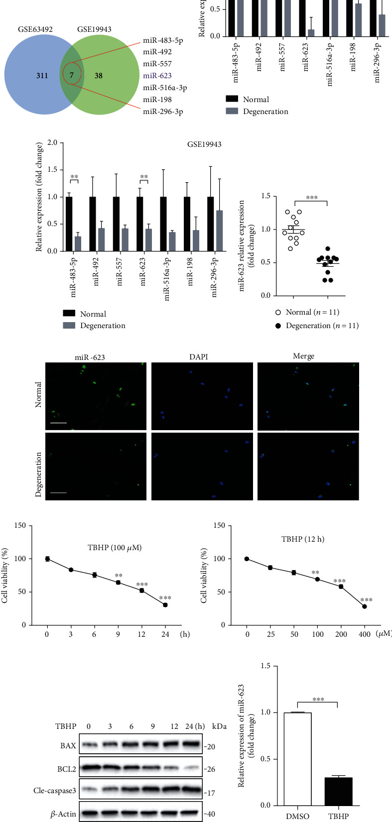
Figure 1.

Oxidative stress dampens miR-623 expression in NP cells. (a) Differentially expressed miRNAs between normal and degenerated NP tissues were determined by analysis with microarray datasets of GSE63492 and GSE19943. Seven miRNAs were found to be downregulated in degenerated NP tissues both in the datasets of GSE63492 and GSE19943. (b) The fold change of relative mRNA expression of seven downregulated miRNAs (a) analyzed by the dataset of GSE63492 was shown as the column chart. (c) The fold change of relative mRNA expression of seven downregulated miRNAs (a) analyzed by the dataset of GSE19943 was shown as the column chart. (d) Quantification analysis of miR-623 mRNA expression in normal NP tissues (n = 11) and degenerated NP tissues (n = 11). (e) Immunostaining analysis of miR-623 mRNA expression in normal NP tissues and degenerated NP tissues. Scale bars represent 50 μm. (f) The survival rate of NP cells incubated with 100 μM TBHP for different hours. (g) The survival rate of NP cells incubated with different doses of TBHP for 12 hours. (h) The protein of BAX, BCL2, and cleaved caspase-3 expression in NP cells induced by 100 μM TBHP for different hours. (i) Quantification analysis of miR-623 mRNA expression in NP cells with or without TBHP stimulation. ∗P < 0.05, ∗∗P < 0.01, and ∗∗∗P < 0.001. P values were analyzed by two-tailed t-tests in (b, c, d, and i) and one-way ANOVA in (f, g).
