FIGURE 2.
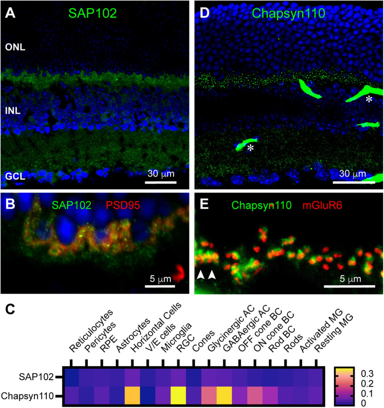
Localization of synaptic scaffolds SAP102 and Chapsyn110 in the mouse retina. (A) Immunostaining for SAP102 (green) with DAPI counterstaining (blue). Labels for retinal nuclear layers are as in Figure 1. SAP102 displayed partially punctate and partially diffuse labeling throughout both inner and outer plexiform layers. Five μm confocal stack. (B) Higher magnification view of the OPL labeled for SAP102 (green) and PSD95 (red). Two μm confocal stack. (C) Average mRNA expression levels of SAP102 (Dlg3) and Chapsyn110 (Dlg2) in mouse retina from single-cell transcriptome data. SAP102 displayed low levels of expression in horizontal cells and Müller glia, and lower levels in rods and cones. Chapsyn110 displayed prominent expression in horizontal cells and some expression in bipolar cells, particularly ON types. Data adapted from Hoang et al. (2020). (D) Immunostaining for Chapsyn110 revealed a sharply punctate distribution in both the outer and inner plexiform layers. Retinal layers are aligned as in (A). Several prominent blood vessels non-specifically labeled by the secondary antibody are present, with two denoted by asterisks. Five μm confocal stack. (E) Higher magnification view of the OPL labeled for Chapsyn110 (green) and mGluR6 (red), labeling the tips of On bipolar cell dendrites. Chapsyn110 aligned closely with mGluR6, but did not co-localize with it. Arrowheads point to clusters of labeling for both markers at a cone pedicle. Three μm confocal stack.

