FIGURE 3.
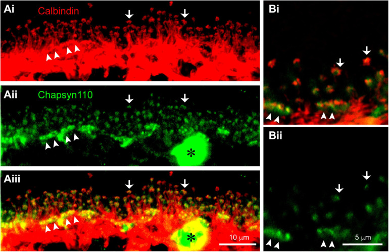
Association of scaffold Chapsyn110 with mouse horizontal cells. (Ai) Immunostaining for Calbindin (red) labels horizontal cells in the OPL. Fine processes rising up are axon terminal processes contacting a single rod at hook-like ending (arrows). Arrowheads point to clusters of dendritic processes contacting cone pedicles. Five μm confocal stack. (Aii) Chapsyn110 immunostaining (green) shows dense clusters of strong labeling (arrowheads) corresponding to locations of cone pedicles and small, isolated clusters of labeling corresponding to the tips of horizontal cell axon terminal processes (arrows). A blood vessel non-specifically labeled by the secondary antibody is marked with an asterisk. (Aiii) Merged view of Calbindin and Chapsyn110 labels reveals co-localization at tips of horizontal cell processes contacting both rods and cones. (Bi) Higher magnification view of horizontal cell processes contacting rods and cones. Labeling scheme is the same as in (A). (Bii) Chapsyn110 labeling in isolation. Chapsyn110 clustered in direct association with horizontal cell process tips, but also in adjacent non-horizontal cell spaces, most likely representing tips of ON-type rod and cone bipolar cells. 0.5 μm confocal stack.

