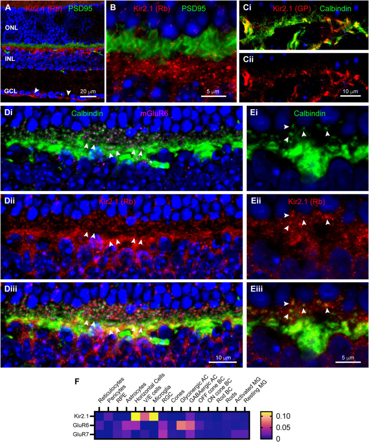
FIGURE 4.
Localization of inward rectifier potassium channel Kir2.1 in mouse retina. (A) Immunostaining for Kir2.1 (rabbit antibody; red) with DAPI counterstaining (blue) and PSD95 (green) to label photoreceptor terminals. Labels for retinal nuclear layers are as in Figure 1. Kir2.1 is diffusely present in the IPL, but strongly labeled at the inner limiting membrane (arrowheads) and in the OPL. Five μm confocal stack. (B) Higher magnification view of the OPL reveals Kir2.1 (red) labeling largely below the photoreceptor terminals (green; PSD95 labeling), but with some diffuse and punctate labeling among the terminals. Two μm confocal stack. (Ci) Kir2.1 labeling (guinea pig antibody; red) colocalizes with Calbindin labeled horizontal cells (green) in the OPL. (Cii) Kir2.1 labeling in isolation. (Di) Another view of horizontal cells in the OPL (Calbindin labeling; green) along with mGluR6 labeling (magenta) to show locations of On bipolar cell dendritic tips. Clusters of mGluR6 label indicate cone terminals; three examples are indicated with paired arrowheads. Six μm confocal stack. (Dii) Kir2.1 labeling (rabbit antibody; red) in the same section. (Diii) Merged view of all three labels. Kir2.1 shows both diffuse and punctate labeling in the vicinity of horizontal cell axon terminal projections contacting rods, but little labeling near clusters of dendritic processes contacting cones. (Ei) Higher magnification view of a horizontal cell labeled with Calbindin antibody (green). Several representative axon terminal tips are highlighted with arrowheads. One μm confocal stack. (Eii) Kir2.1 labeling in the same section. (Eiii) Merged view of the two labels shows that tips of horizontal cell axon terminal processes contain punctate clusters of Kir2.1 labeling (arrowheads). (F) Average mRNA expression levels of Kir2.1 (Kcnj2) and kainate receptor subunits GluR6 (Grik2) and GluR7 (Grik3) in mouse retina from single-cell transcriptome data. Kir2.1 is most prominently expressed in horizontal cells and essentially absent from Müller glia. The kainate receptor subunit GluR6 is also found in horizontal cells and Off bipolar cells, but GluR7 is absent from these cell types. Data adapted from Hoang et al. (2020).

