Figure 2.

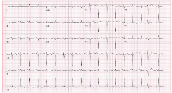
ECG after 6 hours from the presentation showing new T-wave inversions the in lateral precordial leads V5–V6. aVF, Augmented vector foot; aVL, Augmented vector left.

ECG after 6 hours from the presentation showing new T-wave inversions the in lateral precordial leads V5–V6. aVF, Augmented vector foot; aVL, Augmented vector left.