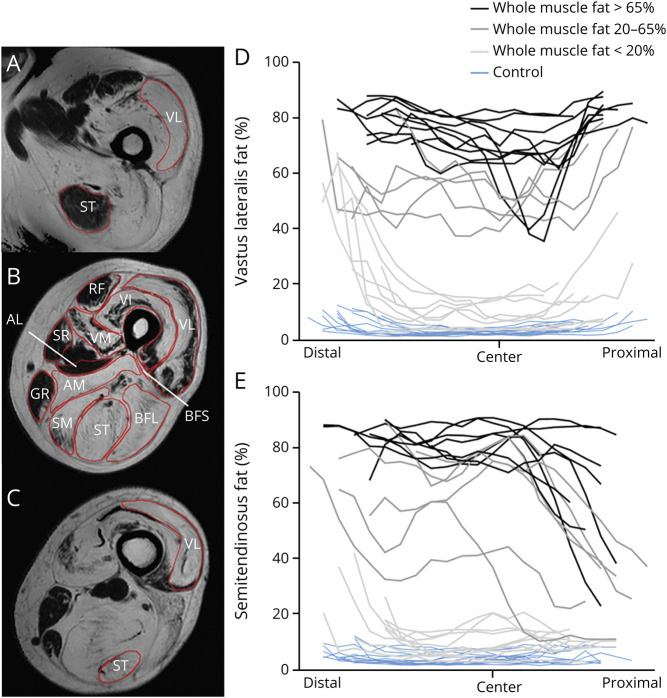
Figure 1. Heterogenous Fat Distribution in Vastus Lateralis (VL) and Semitendinosus (ST).
Example of regions of interest (B) and of heterogenous fat distribution from distal (A) to proximal (C) in the VL (A–D) and ST (A–C, E). (B) An example of heterogenous fat distribution in the axial plane of the quadriceps. To enhance visualization of the fat differences along the proximodistal axis of the muscles, patients are grouped according to their weighted fat of the whole muscle (light gray: <20% fat; dark gray: 20%–65% fat; black: >65% fat). The muscles are aligned based on the insertion of the biceps femoris short head (BFS) (marked as “center”) in D and E. Distal and proximal muscle parts are depicted in the left and right part of the figure, respectively. Blue lines: healthy controls; black/grey lines: Becker muscular dystrophy patients. Each line represents one individual participant. AL = adductor longus; AM = adductor magnus; BFL = biceps femoris long head; GR = gracilis; RF = rectus femoris; SM = semimembranosus; SR = sartorius; VI = vastus intermedius; VM = vastus medialis.

