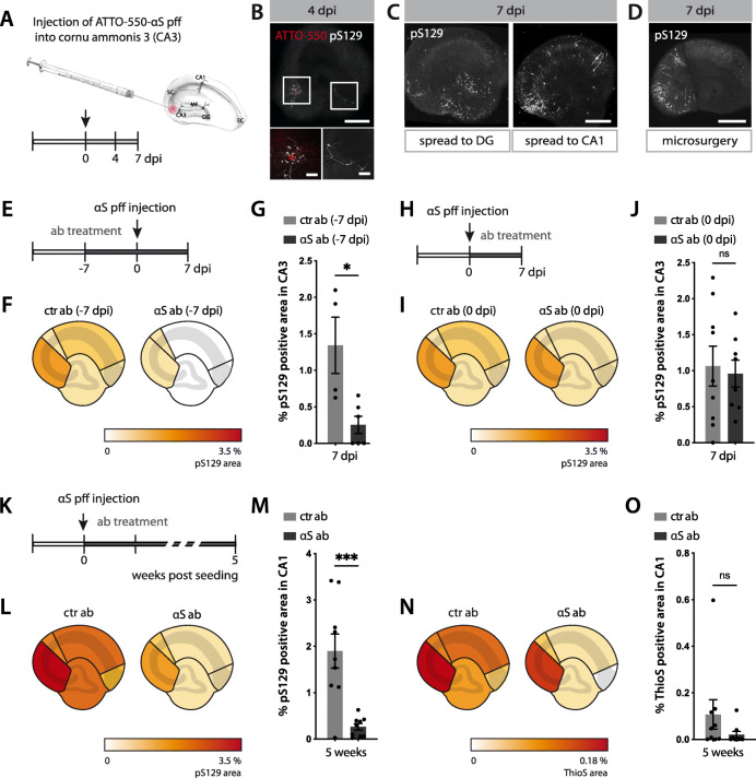
Fig. 4.
Spreading of αS lesions in mouse hippocampal slice cultures. (A) Schematic illustration of the local injection paradigm of ATTO-550-labelled αS pff (from the 35 μM stock solution) into hippocampal CA3 region. (B) ATTO-550-αS pff (red) and the first neuritic pS129-positive inclusions (white) at the injection site (CA3) and in the dentate gyrus (DG) at 4 dpi. Note that αS pff seeds are not phosphorylated upon uptake [32, 39] and therefore the pS129-positive signal can be attributed to aggregated endogenous αS. Scale bars = 500 μm and 100 μm (inserts). (C) Apart from the injection site (CA3) and DG, pS129-positive inclusions also appeared in CA1 at 7 dpi. Scale bars = 500 μm. (D) Disconnecting CA3 from both CA1 and DG via microsurgery before injecting pff inhibited the spreading of the pathology at 7 dpi. Scale bars = 500 μm. (E-G) Antibody-mediated blocking of αS lesion induction. Schematic illustration of experimental setup (E). Seven days prior to αS pff injection into CA3, anti-αS or ctr antibodies were added to the medium (reaching a final concentration of 350 nM) and antibody treatment was continued until 7 dpi. Heatmap of pS129-positive area revealed largely reduced pS129-positive inclusions (F). Quantification of pS129-positive inclusions in CA3 (G). Mean ± SEM; n = 6 and 4 HSCs/group for the αS and ctr antibody, respectively (2 HSCs from ctr group were excluded due to accidental injection into CA1 region); unpaired two-tailed t-test (t(8) = 3.211, p = 0.0124, *p < 0.05). (H-J) Delayed antibody-treatment fails to block αS lesion induction. Schematic illustration of experimental setup (H). Antibodies were added to the culture medium 1 h after αS pff injection into CA3 and antibody treatment was continued until 7 dpi. Heatmap revealed that such delayed antibody treatment did not prevent pS129-positive inclusions in injection region (I). Quantification of pS129-positive inclusions in CA3. Mean ± SEM; n = 9 HSCs/group; unpaired two-tailed t-test (t(14) = 0.7102, p = 0.4892). (K-O) Antibody-mediated blocking of the spreading of the αS lesion. Schematic illustration of experimental setup (K). Antibodies were applied 1 h after αS pff injection into CA3 and antibodies were continuously added until 5 weeks post-seeding. Heatmap revealed that spreading of pS129-positive inclusions to other subregions has been largely reduced (L) and the same was true for ThioS-positive inclusions (that start to develop 2–3 weeks after the neuronal inclusions, see Fig. 2) (N). Quantification of pS129-positive inclusions (M) and ThioS-positive inclusions (O) in CA1. Mean ± SEM; n = 9 HSCs/group; for pS129 (unpaired two-tailed t-test (t(16) = 4.362, ***p = 0.0005); for ThioS, two-tailed Mann-Whitney test (U = 25, p = 0.1782)

