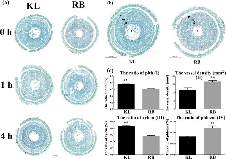
Fig. 1.
Microscopy observations of kiwifruit shoot anatomical structure. (a) Images of the anatomical structure of shoots after 0 h, 1 h, and 4 h of treatment (− 25 °C) for both genotypes. (b) Anatomical structure images of untreated shoots of both genotypes. I, the ratio of pith; II, the vessel density; III, the ratio of xylem; IV, the ratio of phloem. (c) Parameters of the anatomical structure of both genotypes. The bars represent the standard errors of the means (n = 3). The asterisks indicate that the values are significantly different (** for P < 0.01)

