Figure 5.

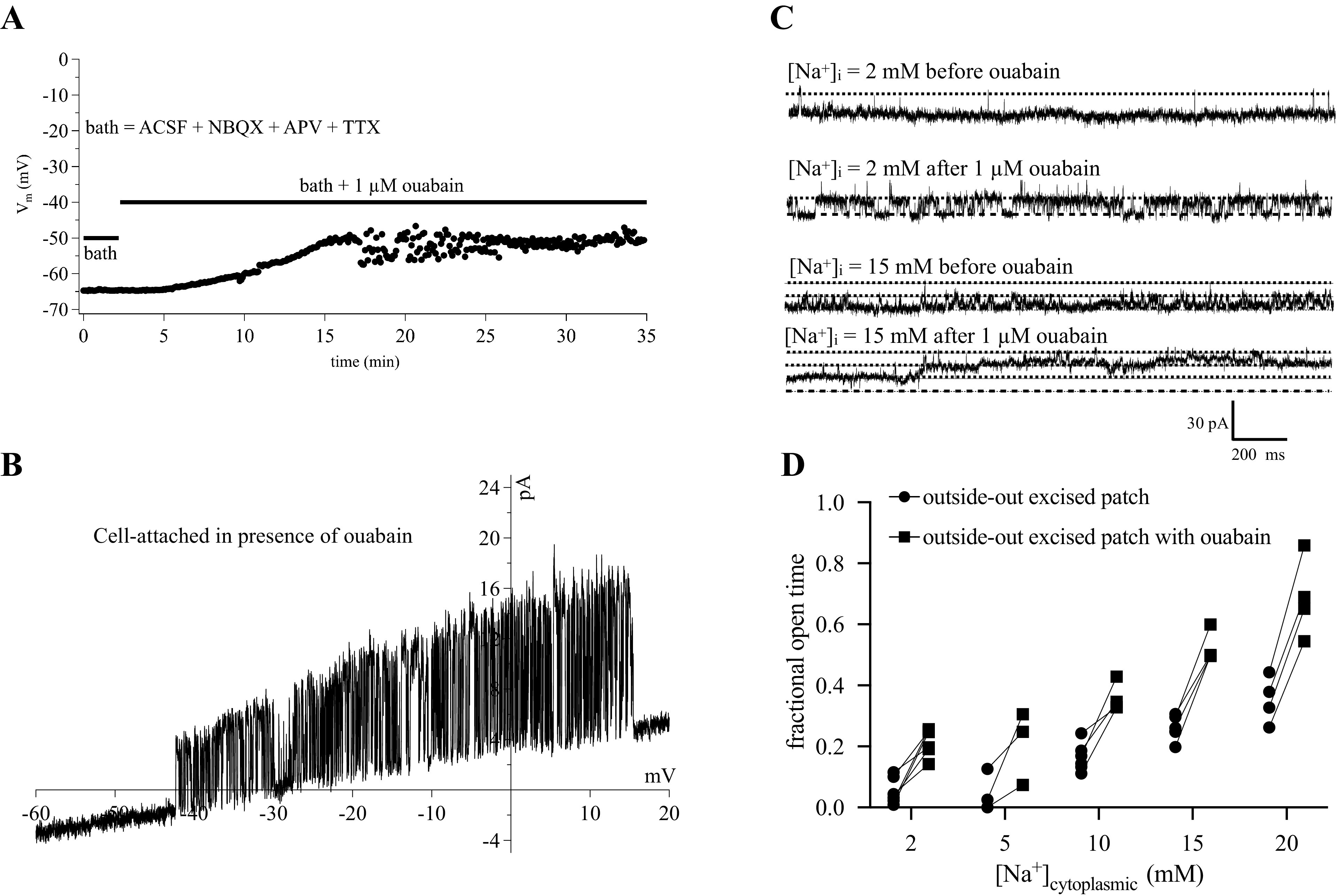
Effects of ouabain. A: a current-clamp recording shows the cell to depolarize to −50 mV in the presence of ouabain (N = 1 cell). B: a cell-attached ramp shows a high degree of activity of a KNa channel (N = 1 cell). Recordings of outside-out excised patches with various concentrations of cytoplasmic Na+. C: example traces before and after application of 1 µM ouabain. D: summary of all experiments from OSO patches before and after ouabain. Repeated measures two-way ANOVA indicated ouabain application increased fractional open time at all Na+ concentrations [F(1,10) = 115.1, P = 0.0001]. ACSF, artificial cerebral spinal fluid; KNa, sodium-activated potassium channels; [Na+]i, intracellular sodium concentration; OSO, outside-out.
