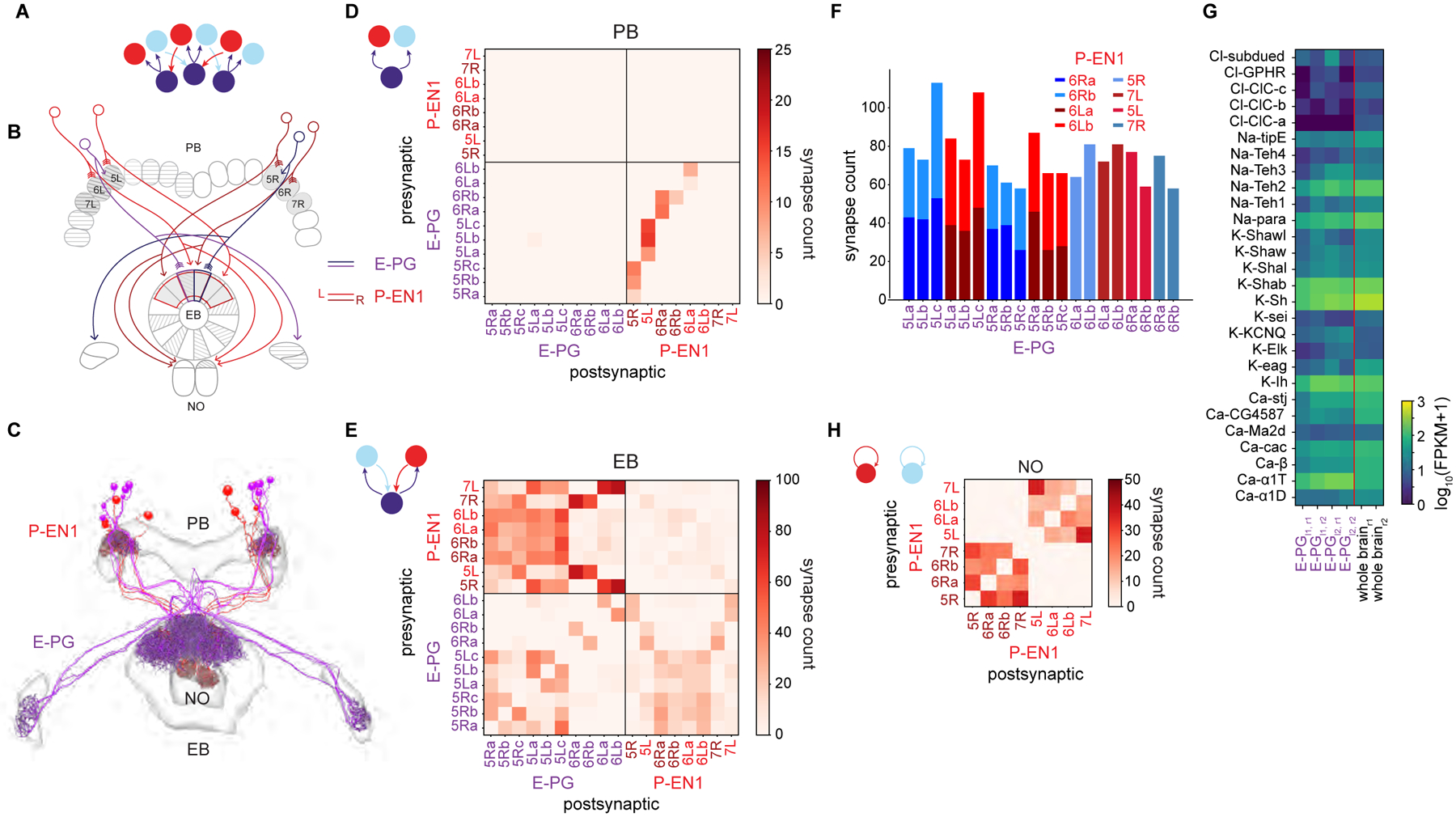
Figure 5: Recurrent loop between E-PGs and P-EN1s that updates bump position during turns is angularly shifted in the EB.

A. Neurons that conjointly encode the fly’s angular velocity and heading (red, light blue) move the bump (purple) around the ring (see Figure 1B).
B. Schematic displaying the anatomically-shifted recurrent loop between sample E-PGs and P-EN1s. EB wedges and PB glomeruli selected for E-PG and P-EN1 reconstruction (Figure 4C) are in gray.
C. E-PG and P-EN1 neurons were manually traced and their synapses onto each other annotated.
D. Connectivity matrix between the E-PGs and the P-EN1s in the PB. The E-PGs synapse onto the P-EN1s that arborize in the same glomerulus (lower right quadrant).
E. Connectivity matrix between the E-PGs and the P-EN 1s in the EB. The shifted P-EN 1s synapse onto the E-PGs (top left quadrant, e.g. P-EN1 6R→E-PG 5L). Note that the E-PGs also synapse onto the P-EN1s in the EB (bottom right quadrant).
F. The total number of synapses from P-EN1s onto individual E-PGs. Although some individual E-PGs receive inputs from single P-EN1s and others from two P-EN1s, the total number of synapses is approximately maintained (p = 0.30, two sample t-test, n = 12 samples for two P-EN1s, n = 8 samples for one P-EN1s).
G. mRNA expression of voltage-gated channels in the E-PGs.
H. Connectivity matrix between P-EN 1s in the NO. Note that P-EN 1s from the same side of the PB are heavily interconnected in the NO.
