
FIGURE 1.
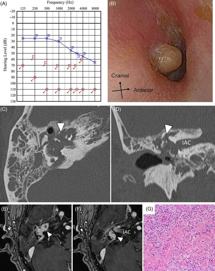
Case 1: A, PTA showed no measurable hearing on the right. B, On observation of the right tympanic membrane, a pulsatile protruding lesion is seen from the tympanic cavity. C,D, CT shows a mass lesion extending from the inner ear to the internal auditory canal, tympanic cavity, and external ear canal (arrowhead in C and D). It is accompanied by the destruction of bones (C: axial image, D: coronal Image). E,F, Contrast‐enhanced MRI revealing an avidly enhancing lesion extending around the inner ear (arrowhead in E and F). The longest diameter of the tumor was 20 mm. G, There is hyperinflammatory granulation tissue growth and fibrosis. The overall picture is consistent with nonspecific inflammation, with no obvious neoplastic lesions observed. CT, computed tomography; IAC, internal auditory canal; MRI, magnetic resonance imaging; PTA, pure tone audiogram