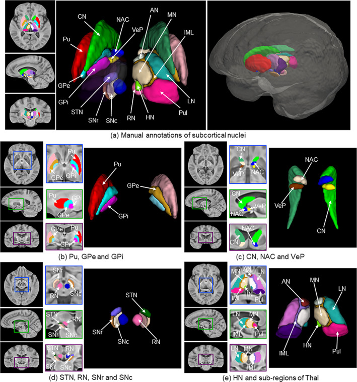
FIGURE 8.

Manual annotations of subcortical brain nuclei. (a) Labeled segmentations overlaid on the sections of the axial, sagittal, and coronal views of the Hybrid template, as well as a 3D rendering of the annotations with the labels of subcortical structures; (b) Labeled Pu, GPe, and GPi on the sections of three views and the 3D rendering; (c) Labeled CN, NAC, and VeP on the sections of three views and the 3D rendering; (d) Labeled STN, RN, SNr and SNc on the sections of three views and the 3D rendering; (e) Labeled HN and five sub‐regions of Thal on the sections of three views and the 3D rendering
