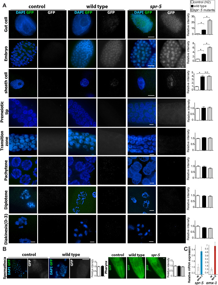
Fig 3. AMX-1 is expressed in the nuclei of gut, embryonic and sheath cells and is further upregulated in absence of SPR-5.
(A) Immunolocalization of AMX-1-GFP utilizing an anti-GFP antibody in the indicated mitotic cells and at different stages in the gonads of control (N2 worms), wild type (amx-1::GFP worms) and spr-5; amx-1::GFP worms. AMX-1-GFP signal is detected in the nuclei of mitotic cells including gut, embryonic and sheath cells. The absence of SPR-5 expression results in increased AMX-1 signal in mitotic cells. Right, relative intensity of AMX-1 expression measurements. *P<0.0001 by the two-tailed Mann–Whitney test, 95% C.I. No distinct signal is detected in either amx-1::GFP or spr-5;amx-1::GFP worms during meiotic progression (Transition zone, Pachytene, Diplotene and Diakinesis). Bars, 10μm. (B) At spermatheca or pharynx, no distinct signal is detected in either amx-1::GFP or spr-5;amx-1::GFP worms. Bars, 10 μm. (C) mRNA expression levels of spr-5 and amx-1 quantified by qRT-PCR. Relative mRNA expression measured in wild type, amx-1, and spr-5 mutants. ~3.2-fold induction of spr-5 mRNA in amx-1 and ~5.8-fold induction of amx-1 mRNA expression in spr-5 mutants are detected.

