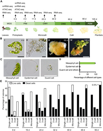
Fig. 1. Regeneration of plants from Arabidopsis mesophyll protoplasts.

(A) Schematic outline of the protoplast regeneration protocol. Time points of sample collection for genome-wide assays are specified. (B) Key stages of mesophyll protoplast regeneration, showing a cell at 4 days after protoplast isolation, a microcallus at 40 days, a callus at 69 days, and a plantlet at 105 days. Scale bars, 25 μm (I) and (II), 1 mm (III), or 1 cm (IV). (C) Protoplasts derived from three cell types after an overnight enzymatic digestion. Scale bar, 25 μm. (D) Percentage of protoplasts derived from mesophyll cells, epidermal cells, guard cells, and other cell types after an overnight enzymatic digestion (182 ≤ n ≤ 204). Data are presented as means ± SD for more than three independent experiments. (E) Distribution of mesophyll protoplast cells during regeneration, measured at various time points after protoplast isolation (n = 376).
