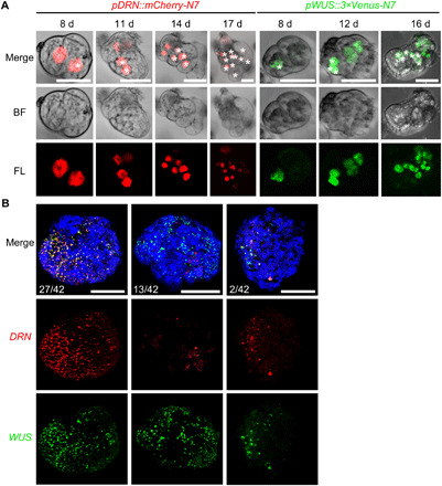
Fig. 6. WUS and DRN are maintained in regeneration microcalli and calli.

(A) Time-lapse images showing the maintained expression of pDRN::mCherry-N7 (red) and pWUS::3×Venus-N7 (green) in the regenerating mesophyll protoplasts. Asterisks label progenitors of the same cells in corresponding time points. Scale bars, 25 μm. (B) Maximum intensity views of confocal images showing the coexpression of pDRN::mCherry-N7 (red) and pWUS::3×Venus-N7 (green) in representative regenerating calli at 60 days. Three expression patterns are shown. Scale bars, 250 μm.
