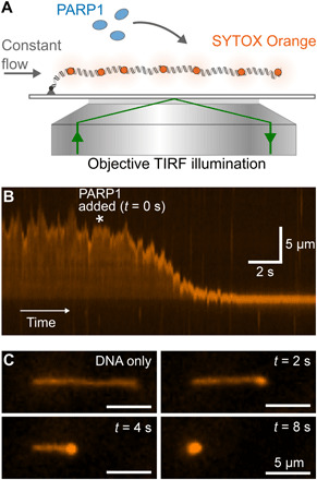
Fig. 1. TIRF imaging shows condensation of DNA by PARP1.

(A) Schematic of TIRF microscopy imaging of a single λ-DNA molecule stained with SYTOX Orange. A constant flow was maintained, which stretches out the DNA over a distance close to its contour length. (B) Kymograph showing DNA extension over time. 400 nM PARP1 is added at the time point indicated by the asterisk. (C) Snapshots showing individual image frames at the indicated time points.
