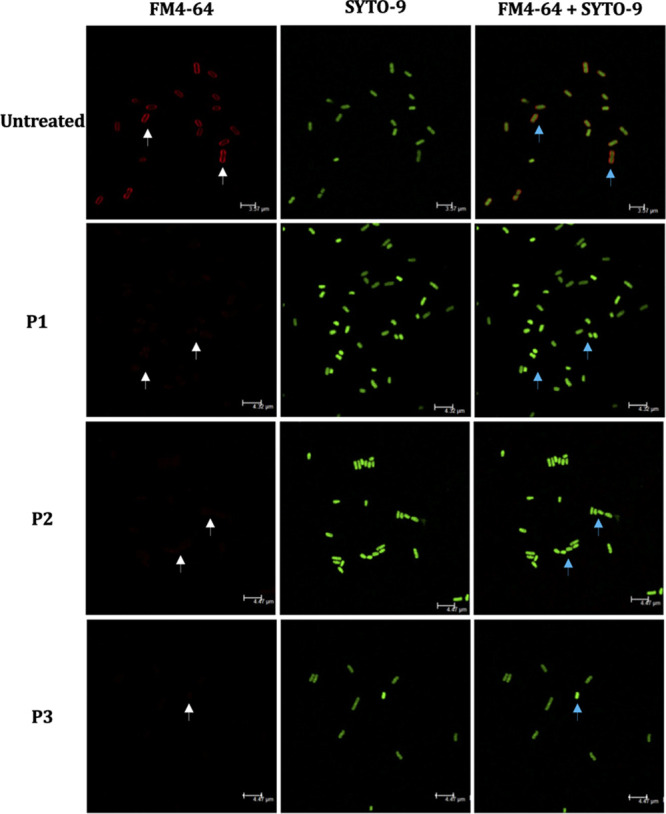
FIG 3.

Confocal fluorescence images of APEC either untreated or treated with peptides. Red, FM4-64 (membrane stain); green, SYTO-9 (nuclear stain). APEC O78 cultures were treated with peptides (5× MIC), incubated (3 h), stained with FM4-64 and SYTO-9 (45 min), and imaged using a Leica TCS SP6 confocal scanning microscope. APEC membrane was clearly visible in untreated APEC (white arrows), whereas no or minimally visible membrane was observed in APEC treated with peptides. Superimposed images (FM4-64 plus SYTO-9) showed nuclear material of APEC enclosed by membrane in untreated APEC (blue arrows), whereas no membrane was visible covering the nuclear material in peptide-treated APEC.
