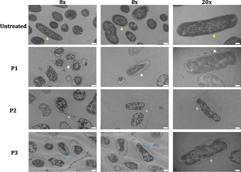
FIG 4.

Transmission electron microscopy images (×8 and ×20 magnifications) of APEC either untreated or treated with peptides. APEC O78 cultures were treated with peptides (10× MIC), incubated (3 h), and imaged using a Hitachi H-7500 microscope. Clearly demarcated membrane encircling the dense cytoplasmic contents was observed in untreated APEC (yellow arrows), whereas the membrane was either sloughed/shed (white arrows), flaccid (orange arrows), or ruptured (blue arrows) in APEC treated with peptides. Bars, 0.2 μm.
