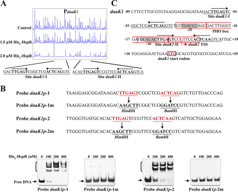
FIG 5.
HspR-binding site on dnaK1 promoter region. (A) DNase I footprinting assay of HspR on dnaK1p. (B) EMSAs using 50-nt WT probes dnaK1p-1 and dnaK1p-2 and their mutated probes dnaK1p-1m and dnaK1p-2m. Each lane contained 0.15 nM labeled probe. (C) Nucleotide sequences of dnaK1 promoter region and HspR-binding sites. Numbers indicate distance (nucleotides) from dnaK1 TSS. Black box, dnaK1 TSC; bent arrow, dnaK1 TSS; red boxes, PHO box. Other notations as in Fig. 3C.

