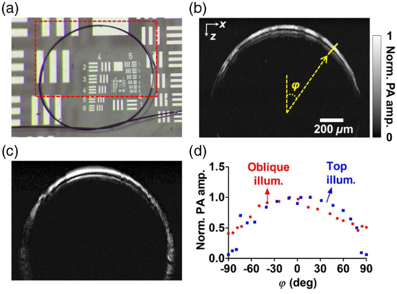
Fig. 5.
(a) A photo of the phantom vessel comprised of five carbon fibers. Each of the fibers is around in diameter. (b)–(c) Images of the phantom in the red dashed region with the top illumination beam and the oblique illumination beam, respectively. (d) Peak PA amplitudes sampled at different angles in (b) and (c), as illustrated by the yellow line in (b).

