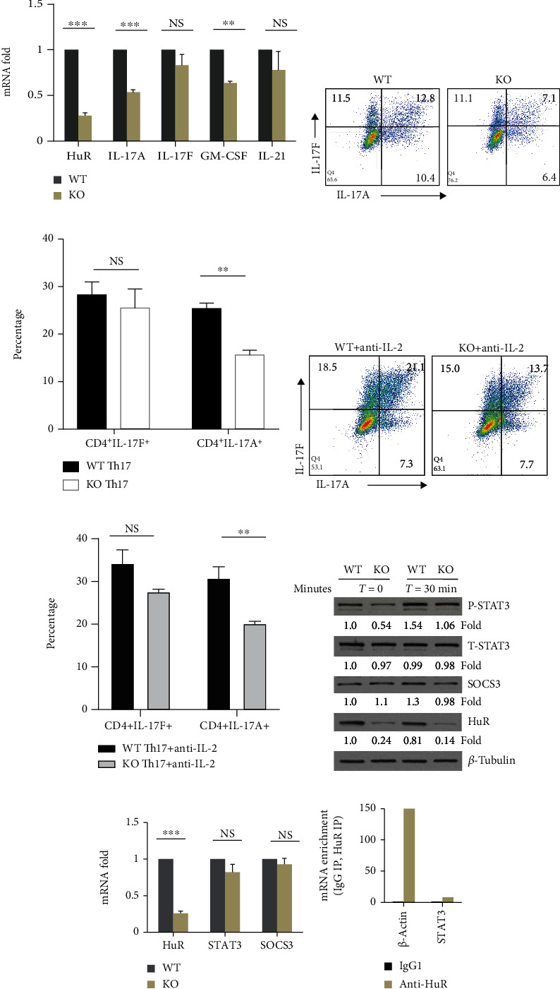
Figure 3.

Genetic deletion of HuR reduces the activities of STAT-3 in Th17 cells. (a) Naïve CD4+ T cells were stimulated under Th17 cell polarization condition for 4-5 days. The levels of cytokine mRNAs were measured by RT-qPCR. (b) Flow cytometry assay showed that percentage of IL-17A-positive cells significantly decreased, but that of IL-17F-positive cells slightly decreased. (c) Summary of flow data for Th17 cell culture from three independent experiments (mean ± SEM). (d) WT and HuR KO Th17 cells were cultured under Th17 cell culture condition with anti-IL-2 (30 μg/ml) for 5 days. Representative flow cytometric data was shown. (e) Summary of flow data for Th17 cell culture with or without anti-IL-2 from three independent experiments (mean ± SEM). (f) Th17 cells were rested 2 days in the presence of IL-2 (3 ng/ml) following restimulation in the presence of Th17 cell-polarizing cytokines (TGF-β+IL-6) without IL-23 for the indicated times. Western blot assay showed that HuR deficiency in Th17 cells reduced the level of p-STAT3 compared to WT Th17 cells, but not total STAT-3. (g) Knockout of HuR did not reduce the level of Stat3 and SOCS3 mRNA in Th17 cells as characterized by RT-qPCR. (h) RIP assay indicated that HuR did not bind to Stat3 mRNA. Data in (a), (c), (e), and (g) represents the summary of three independent experiments. Student's t-test was used for statistics analysis. ∗p < 0.05, ∗∗p < 0.01, and ∗∗∗p < 0.001. Data in (b), (d), (f), and (h) represent one of the three independent experiments.
