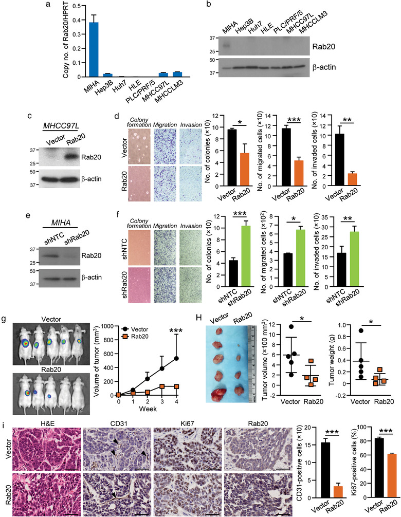
FIGURE 2.

Rab20 negatively regulates growth, motility and tumorigenic potentials of HCC cells. (a‐b) mRNA and protein levels of Rab20 in MIHA and HCC cell lines were quantified by quantitative RT‐PCR and immunoblotting, respectively. (c) MHCC97L was stably transduced with Rab20 expression plasmid and empty vector, and immunoblotted with anti‐Rab20 and anti‐β‐actin antibodies. (d) MHCC97L Vector and Rab20 cells were subjected to soft agar (Left), migration (Middle) and invasion (Right) assays. Number of colonies, migrated and invaded cells were counted. Representative images of colonies and cells are shown. (e) Immunoblot image showing the expression of Rab20 in MIHA stably transduced with non‐targeted shRNA control (shNTC) and shRNA targeting Rab20 (shRab20). (f) MIHA shNTC and shRab20 cells were subjected to soft agar (Left), migration (Middle) and invasion (Right) assays. Number of colonies, migrated and invaded cells were counted. Representative images of colonies and cells are shown. (g) MHCC97L Vector and Rab20 cells were subcutaneously injected into nude mice. Tumour growth rate was monitored and measured weekly. Four weeks after injection, bioluminescence signals emitted by the subcutaneous tumours were measured. (h) Volume and size of the excised xenograft were recorded and compared. (i) Paraffin‐embedded tissues obtained from xenograft were stained with H&E, anti‐CD31, anti‐Ki67 and anti‐Rab20 antibodies. Arrow indicates the presence of stained blood vessels. The numbers of CD31‐ and Ki67‐positive cells were quantified and plotted. Scale bar: 100 μm. Data are represented as mean ± SEM. *P < 0.05, **P < 0.01, ***P < 0.001. P < 0.05 is considered as statistically significant
