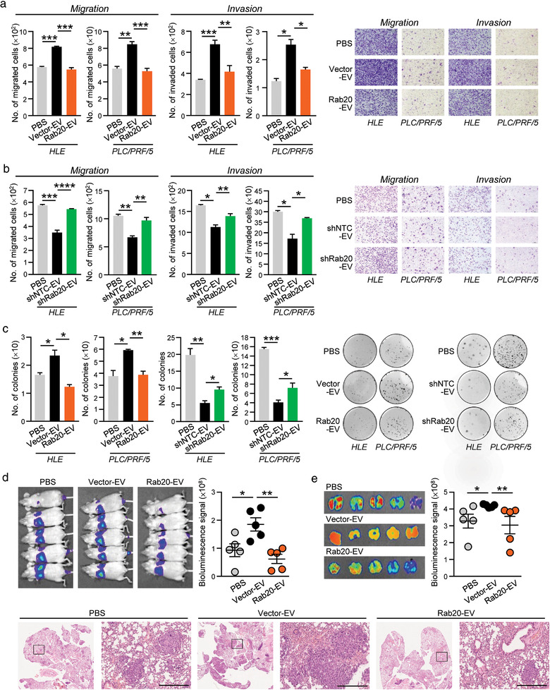
FIGURE 3.

Diminished promoting activity of EVs released by MHCC97L cells with Rab20 overexpression. HLE and PLC/PRF/5 cells were treated with MHCC97L Vector‐ and Rab20‐EV (a), and MIHA shNTC‐ and shRab20‐EV (b). Treated HCC cells were seeded for migration and invasion assays. Representative images were shown and the number of cells was quantified. (c) Colony formation assay was performed on HLE and PLC/PRF/5 cells treated with the indicated EVs. Colonies formed were fixed, stained and counted. (d) p53‐/‐;Myc‐transduced mouse hepatoblasts were co‐injected with PBS, MHCC97L Vector‐ and Rab20‐EV in nude mice through tail vein. Two weeks after co‐injection, bioluminescence imaging of animals was performed. (e) Ex vivo bioluminescence imaging of the excised lungs was performed. Representative H&E‐stained images showing tumour nodules in the lungs were shown. Scale bar: 500 μm. Data are represented as mean ± SEM. *P < 0.05, **P < 0.01, ***P < 0.001, ****P < 0.0001. P < 0.05 is considered as statistically significant
