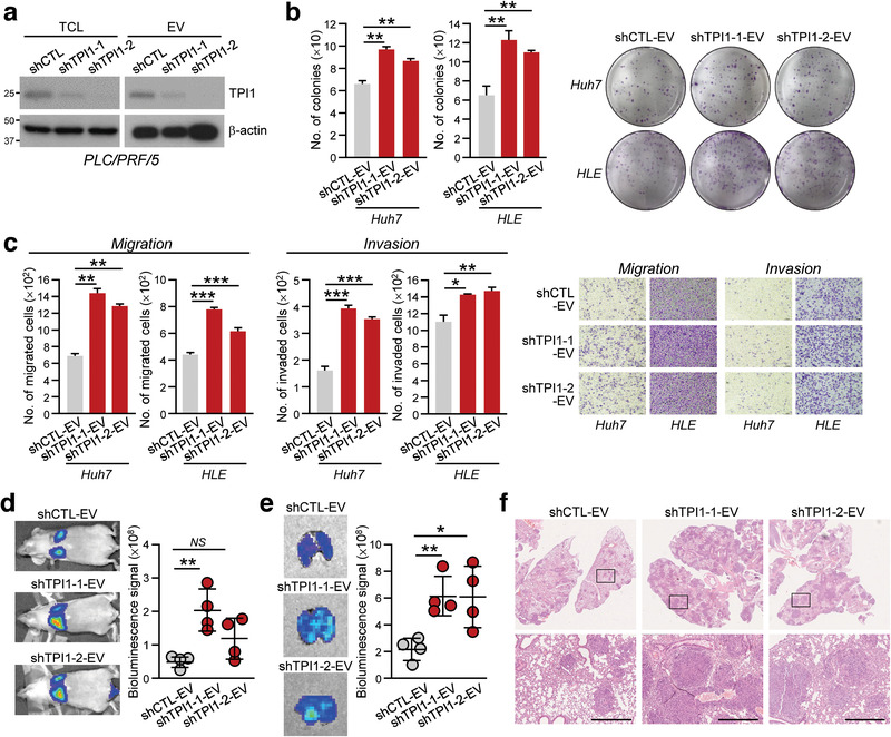
FIGURE 5.

EVs with reduced TPI1 expression promoted aggressiveness of HCC cells. (a) PLC/PRF/5 cells were stably transduced with control shRNA (shCTL) and shRNA targeting TPI1 (shTPI1‐1 and shTPI1‐2). Total cell lysates (TCL) and isolated EVs were immunoblotted with anti‐TPI1 and anti‐β‐actin antibodies. (b) Huh7 and HLE treated with shCTL‐, shTPI1‐1 and shTPI1‐2‐EVs were subjected to colony formation assay. (c) Migratory and invasive abilities of Huh7 and HLE cells treated with shCTL‐, shTPI1‐1 and shTPI1‐2‐EVs were determined by migration and invasion assays. (d) Two weeks after co‐injection of p53‐/‐;Myc‐transduced murine hepatoblasts with shCTL‐, shTPI1‐1 and shTPI1‐2‐EVs, mice were subjected to bioluminescence imaging. Representative images of animals of each group are shown. (e) Lungs were excised and subjected to bioluminescence signal detection. (f) Representative H&E‐stained images showing tumour nodules in the lungs were shown. Scale bar: 500 μm. Data are represented as mean ± SEM. *P < 0.05, **P < 0.01, ***P < 0.001. P < 0.05 is considered as statistically significant
