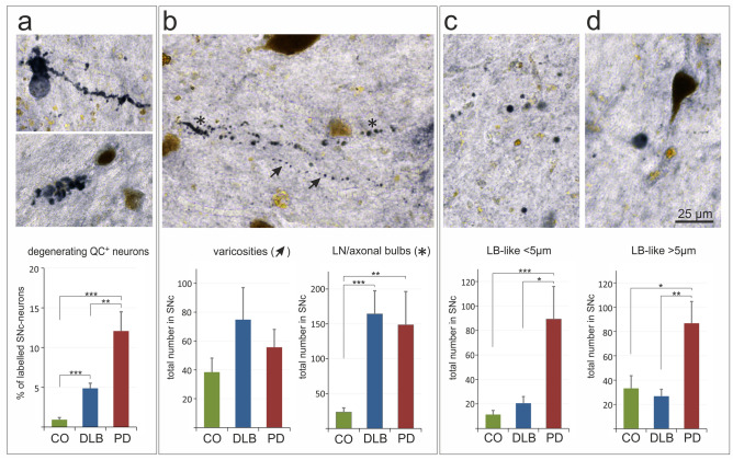
Fig. 6.
QC in human SN: presence in neuropathological structures. QC (labelled in black) was found to be associated with degenerating neurons (a), with varicosities (arrows) and Lewy neurites/axonal bulbs (asterisks) (b), as well as with small (c) and large (d) Lewy body-like structures. The quantification of these structures revealed a high association of QC with Lewy body-like aggregates in PD, but not in DLB (c, d) no significant differences in the number of QC-positive varicosities between groups, but a similarly strong increase in the number of QC immunoreactive Lewy neurites and axonal bulbs in DLB and PD compared to controls (CO) (b), and a more abundant association of QC with degenerating neurons in PD than in DLB (a). Brown color arises from neuromelanin. Mean ± SEM, n = 10, Statistical significance at *p < 0.05; **p < 0.01; ***p < 0.001 defined by t-test

