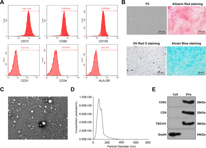
Fig. 1. Identification of HucMSCs and HucMSCs-EVs.
A Surface markers of HucMSCs: CD73, CD90, and CD105 (positive markers), and CD31, HLA-DR, and CD34 (negative markers) were detected using flow cytometry. B Morphology of HucMSCs was observed under a light microscope (left); osteogenesis, adipogenesis, and chondrogenesis were observed using alizarin red staining, oil red O staining, and alcian blue staining respectively. C the ultrastructure of HucMSCs-EVs was observed by TEM. D the particle size of EVs was detected using Nanosight analysis. E the surface marker proteins of EVs (CD63, CD9, and TSG101) were detected using Western blotting. The experiment was repeated three times.

