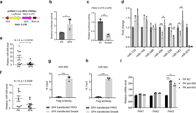
Fig. 5. Smad4 negatively regulates PAK3 via transactivation of miR-495 and miR-543 expression.
a A luciferase reporter fused to PAK3 3′ UTRs was constructed in a pcDNA3.1-Luc-MCS plasmid. b Luciferase activities of PK and SPK cells transfected with PAK3 3′UTR-Luc plasmid confirmed increased PAK3 expression level SPK cells. Data presented are the mean ± SD from three biologically independent samples (n = 3); *p = 0.023816011, as determined by two-tailed Student’s t-test. c Luciferase reporter assay showing that overexpression of Smad4 decreased PAK3 expression level in SPK cells. Data presented are the mean ± SEM from three biologically independent samples (n = 3); ***p = 1.58613E-05, as determined by a two-tailed Student’s t-test. d RT-PCR detected expression of PAK3 correlated miRNAs in PK, SPK, and SPK overexpressing Smad4 cells. Data presented are the mean ± SEM from three biologically independent samples (n = 3); *p = 0.012761254; **p = 0.00456168; △p = 0.005190084; ○p = 0.015974559; ☆p = 0.024995116; as determined by two-tailed Student’s t-test. e, f Expression levels of miR-495 and miR-543 were determined in PK and SPK mouse lung tumors. n = 9, data represent means ± SEM; p < 0.05, as determined by two-tailed Student’s t-test. g, h The interaction between Smad4 and miR-495 (g) or miR-543 (h) in SPK cells were verified by ChIP assay. Data presented are the mean ± SEM from three biologically independent samples (n = 3); ***(h)p = 6.64287E-06; ***(g)p = 0.00021899; as determined by two-tailed Student’s t-test. i qRT-PCR analysis gene expression in PK cells. Data presented are the mean ± SEM from three biologically independent samples (n = 3). ***p = 5.32332E-07; **p = 0.000179381.

