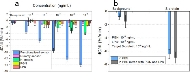
Fig. 4. Specificity verification of the immunosenors.
a Specificity verification using different types of sensors and different analytes. The background is first tested to verify the blank control and the sensor blocking effect. Functionalized sensors are compared with dummy sensors (also blocked with lactalbumin), to characterize the antibody functionalization. The control group contains nucleocapsid (N-) protein, peptidoglycan (PGN), and lipopolysaccharide (LPS). b Specificity verification in the hybrid medium. A hybrid medium is constructed by mixing PGN and LPS in 0.1× PBS solution. The PGN and LPS concentrations in 0.1× PBS are both 10−3 ng/mL, and the spiked S-protein is at 10−4 ng/mL. In both (a, b), the error bar represents the standard deviation from three tests of the same sample using different sensors of the same batch.

