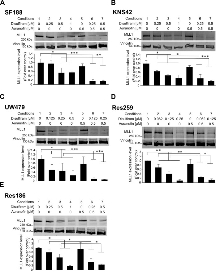
Fig. 2. Disulfiram induces MLL degradation.
A–E Top: whole-cell lysates were prepared from the indicated cell lines, cultured in the absence or presence of 0.5 mM auranofin with increasing concentrations of disulfiram for 16 h. Extracts were probed with α-MLL1 antibody and α-vinculin antibody as loading control. Bottom: the graphs represent image analysis of band intensity. Data are mean ± STD; n = 3; ∗p < 0.05, ∗∗p < 0.01 ∗∗∗p < 0.001; two-way analysis of variance (ANOVA) test with Bonferroni posttest.

