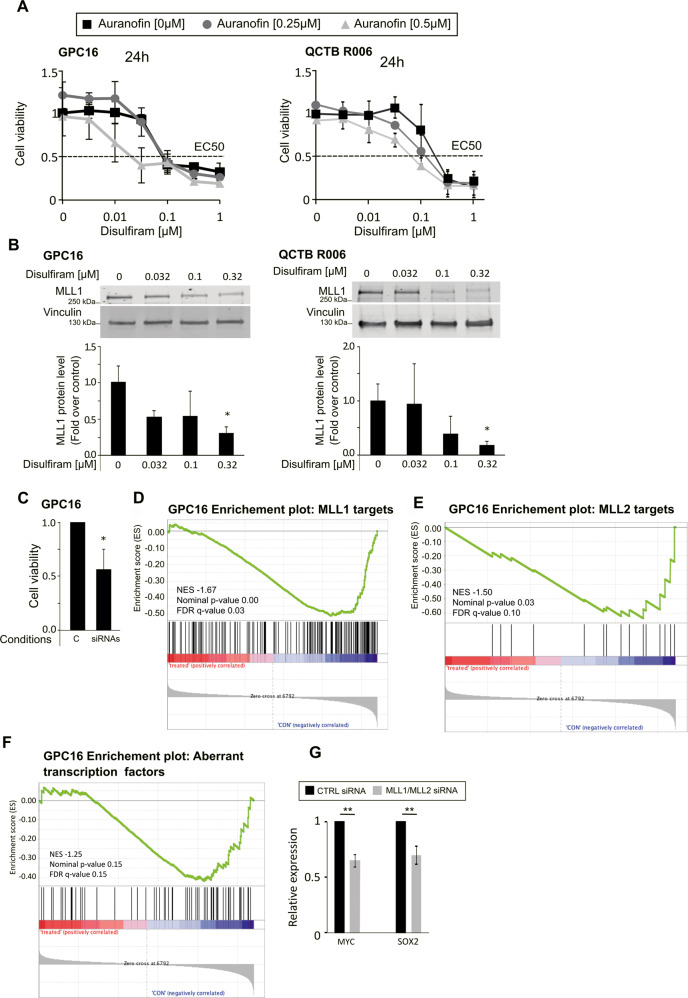
Fig. 5. Disulfiram inhibits cell proliferation and induces MLL degradation in patient-derived primary high-grade glioma cells.
A Cells were plated in 96-wells plates. The following day cells were treated with the indicated concentrations of disulfiram in the absence or presence of increasing doses of auranofin. At the indicated time cell viability assays were performed. Data are mean ± STD; n = 3. B Top: whole-cell lysates were prepared from the indicated cells, cultured in the absence or presence of increasing concentrations of disulfiram for 16 h. Extracts were probed with α-MLL1 antibody and α-vinculin antibody as loading control. Bottom: the graphs represent image analysis of band intensity. Data are mean ± STD; n = 3; ∗p < 0.05; two-way analysis of variance (ANOVA) test with Bonferroni posttest. C GPC16 cells were seeded in six-well plates at a density of 0.4 × 10⁶ cells/well and transfected with 25 pmol ON-TARGETplus Control pool Non-Targeting pool (labeled C), or ON-TARGETplus SMART pool siRNA targeting MLL1 and MLL2 (labeled siRNA). Cell viability assays were performed 3 days after transfection. Data are mean ± STD; n = 3; ∗p < 0.05; one-sample t-test. D, E, and F same experiment as Fig. 1 but with GPC16 cells. G qPCR analyses of selected transcription factors expression upon MLL1/MLL2 downregulation in GPC16 cells. Data are mean ± STD; n = 3; ∗∗p < 0.01; one-sample t-test.

