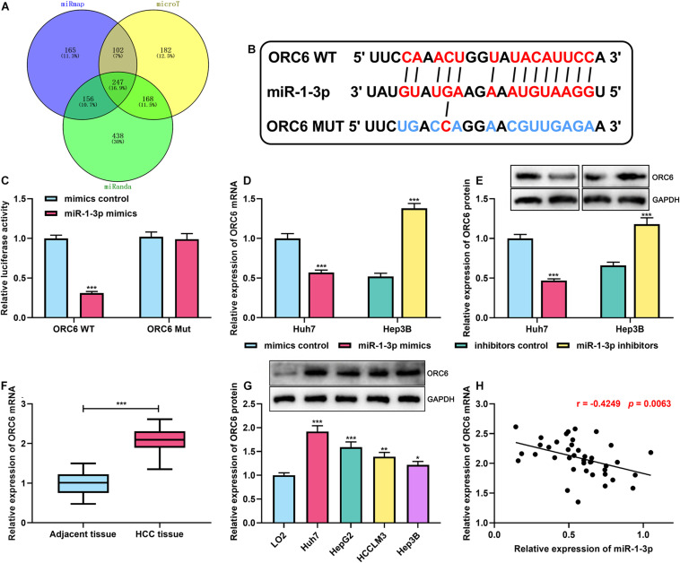
FIGURE 3.
MiR-1-3p directly targeted ORC6. (A) Venn diagram showed overlapped target genes of miR–1–3p in miRmap, microT, and miRanda databases. (B) The binding site between ORC6 mRNA 3′UTR and miR-1-3p was co-predicted by miRmap, microT, and miRanda databases. (C) A dual-luciferase reporter assay was performed to validate the binding site between miR-1-3p and ORC6. (D) The effect of miR-1-3p on ORC6 mRNA expression was measured by qRT-PCR. (E) The effect of miR-1-3p on ORC6 protein expression was detected by Westen blot. (F) Expression of ORC6 in HCC tissues and adjacent liver tissues was examined by qRT-PCR. (G) Expression of ORC6 in normal liver cells and HCC cell lines was examined utilizing Western blot. (H) The correlation between miR-1-3p expression and ORC6 expression in HCC tissues was analyzed. All of the experiments were performed in triplicate. *, **, and *** represent p < 0.05, p < 0.01, and p < 0.001, respectively.

