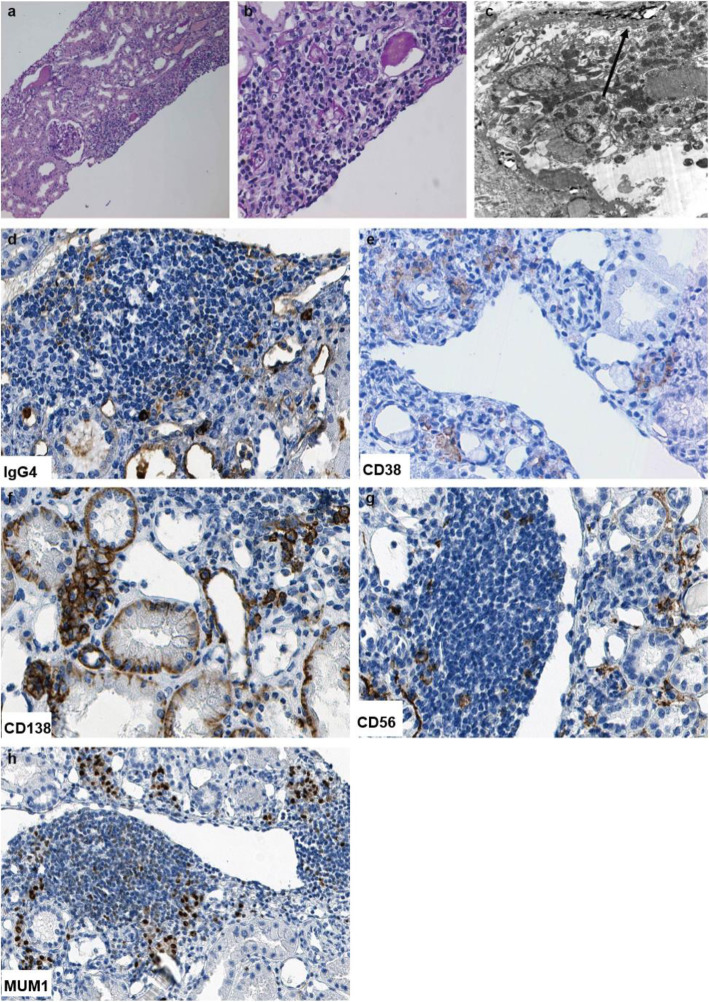
Fig. 5.
Typical pathological features of IgG4-Tubulointersitial nephritis and immunohistochemistry staining of surface biomarkers of plasma cells in renal biopsy. The renal interstitium is infiltrated by plasma cells and lymphocytes predominantly with fibrosis on periodic acid-Schiff (PAS) staining. Original magnification 100x (a) and 400× (b). TBM electron-dense deposits were seen under electron microscope. Original magnification 8000x (c). Marked increase in IgG4-positive plasma cells was seen in the infiltrated cells on immunohistochemistry staining. Original magnification 400× (d). CD38-positive plasma cells (e), CD138-positive plasma cells (f), CD56-positive plasma cells (g), and MUM1-positive plasma cells (h) were seen in the interstitium on immunohistochemistry staining. Original magnification 400 ×

