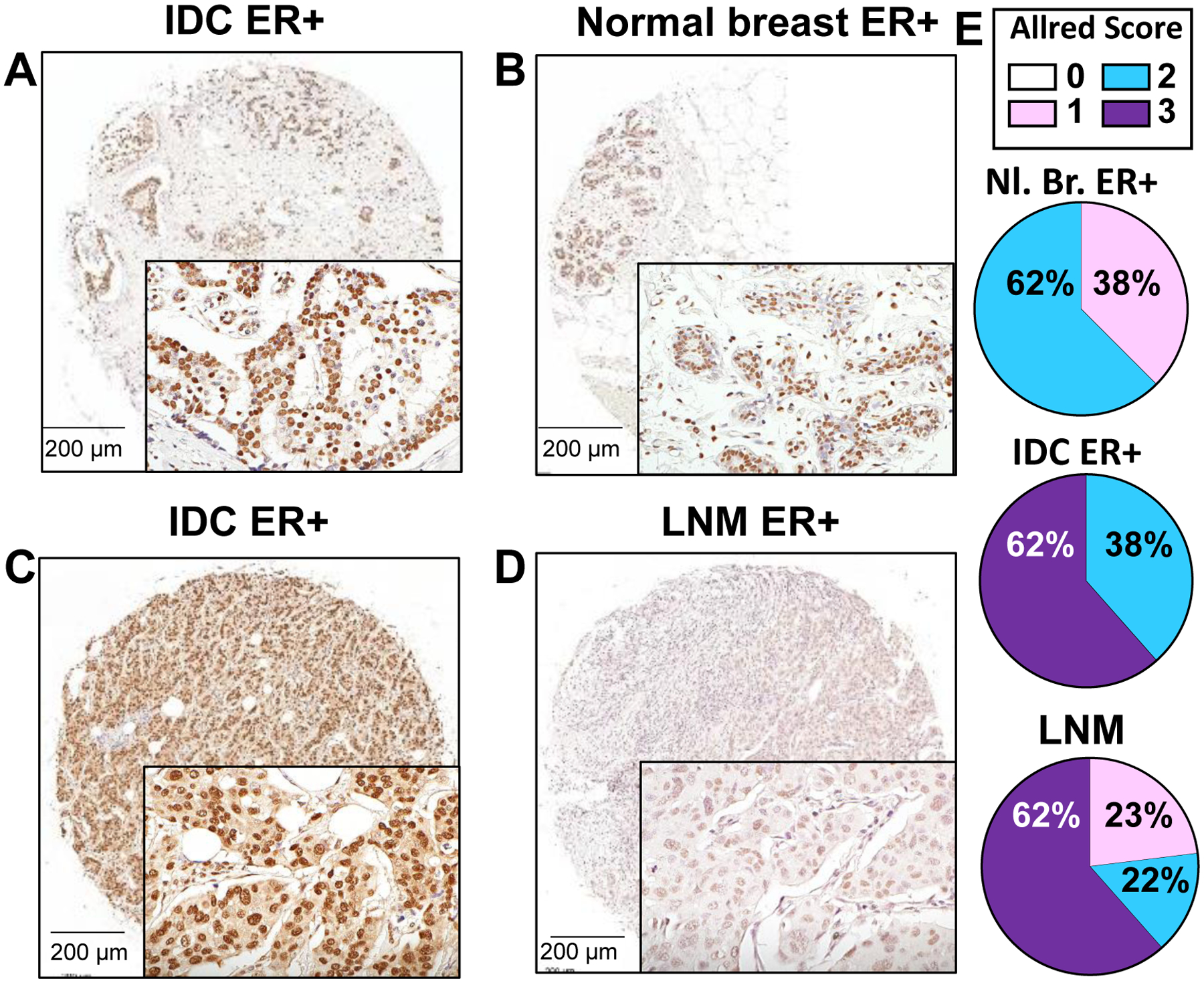
Figure 1: IHC staining for HNRNPA2B1 (A2B1) in ER+ invasive breast carcinoma (IDC) tumors paired with normal breast or lymph node (LN) metastasis from the same patient.

Shown are representative images from a stained TMA. The tumor in A (Allred score 3) is paired with normal breast tissue (B) from the same patient (Allred score 2). The tumor in C (Allred score 3) is paired with its LN metastasis in D (Allred score 1). Bar is 200 μm. Images were taken under 20x magnification. E) Quantification of Allred scores based on intensity and percent of cells stained are indicated as the percent total number of that sample in the TMA (Nl. Breast ER+ = 8; IDC ER+ with matched LNM ER+ = 13).
