FIGURE 3.

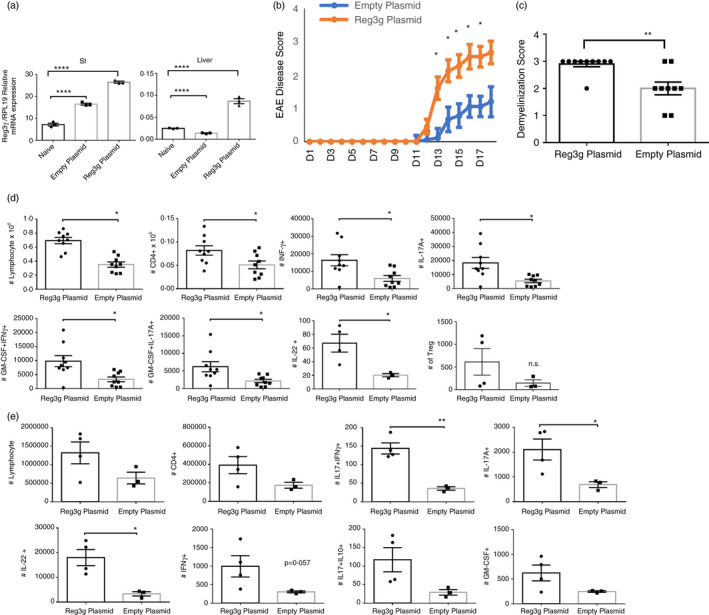
Temporal Reg3γ overexpression via hydrodynamic gene delivery exacerbates experimental autoimmune encephalomyelitis (EAE) pathology and disease scores (a) Reg3γ plasmid or empty plasmid were injected on day −2, and 3 days later, Reg3γ message was quantified by real‐time qPCR in the liver and small intestine tissue (n = 3). (b) Reg3γ plasmid or empty vector was injected on day −2, and EAE was induced by MOG35‐55 immunization on day 0. Mice were monitored for 3‐4 weeks, and EAE disease was scored (n = 9–10 per group). (c) Spinal cords were removed and stained with Luxol fast blue to quantify demyelination 3 weeks after EAE induction (n = 9–10 per group). (d) Spinal cords and brain tissues from Reg3γ plasmid‐ or empty vector‐injected mouse groups were harvested 3 weeks after EAE induction, and infiltrating lymphocytes were quantified based on IFN‐γ, IL‐17A and GM‐CSF production. Absolute number of indicated cytokine‐producing CD4+ T cells or Treg cells (last panel) was quantified (n = 9–10 mice per group). (e) Absolute number of indicated cytokine‐producing CD4+ T cells in the draining LNs of Reg3γ plasmid‐ or empty plasmid‐injected mouse groups at 3 weeks post‐immunization of EAE. (n = 4 mice per group). (*) indicates p‐value <0·05; (**), p < 0·01; (***), p < 0·001; and (****), p < 0·0001
