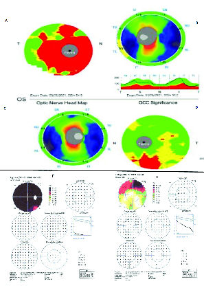
Figure 2.

A and B illustrate ganglion cell complex (GCC) and retinal nerve fiber layer (rNFL) thickness of the right eye, respectively. GCC image demonstrates significant thinning (red area), and rNFL looks normal. C and D show rNFL thickness and GCC of the left eye, respectively. GCC image demonstrates thinning inferiorly (red area)andrNFL looks normal. E and F are Octopus EyeSuite Static perimetry V6.3.1 visual field of the right and left eyes, respectively. Right eye visual field shows severe generalized depression, and the left eye depicts a superior altitudinal defect.
