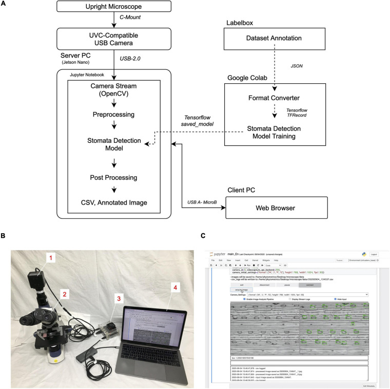
FIGURE 1.
Stomatal detection platform. (A) Schematic diagram of the workflow. (B) Appearance of the platform. Numbers in insets describe the individual components. 1, UVC-compatible camera (ELP-USB13M02-MFV); 2, upright trinocular microscope (SW380T); 3, server PC (Jetson Nano B01); 4, client PC (Macbook Pro 13-inch, 2017). (C) Screenshot of the GUI run in the client PC through the Google Chrome web browser.

