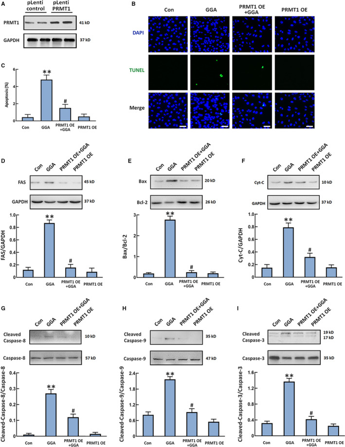
FIGURE 6.

Overexpression of PRMT1 suppresses GGA‐induced U‐2 OS cell apoptosis by inhibiting the FAS‐regulated apoptotic pathway. U‐2 OS cells were transfected with pLenti control or pLenti‐PRMT1 plasmids and treated with 20 µM GGA. A, Western blot detection of PRMT1 expression in U‐2 OS cells. B, Representative photomicrographs of the TUNEL assay (green). Total nuclear staining with DAPI (blue). Scale bar =100 µm. C, The numbers of apoptotic cells were counted in five randomly selected fields for each sample based on the TUNEL images. D‐I, FAS expression (D), the Bax/Bcl‐2 ratio (E), Cyt c release (F), cleaved caspase‐3/caspase‐3 expression (G), cleaved caspase‐8/caspase‐8 expression (H) and cleaved caspase‐9/caspase‐9 expression (I) were detected by Western blot. Quantification of the relative protein levels in the panels under the images. n = 3. **P < 0.01 vs the control group. # P < 0.05 vs the GGA group
