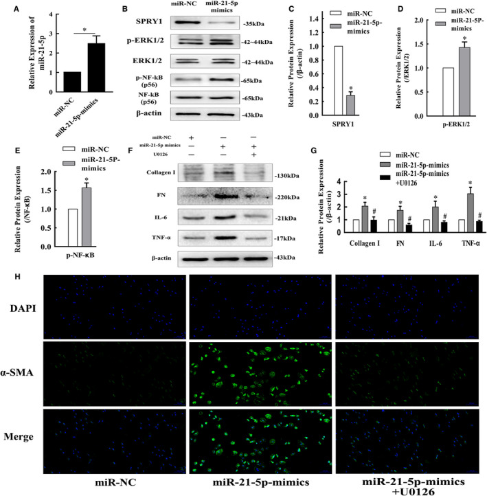
FIGURE 5.

Enhanced miR‐21‐5p expression in HK‐2 cells promoted inflammation and fibrosis development via the SPRY1/ERK/NF‐kB signalling pathway. A, The fold change in miR‐21‐5p levels in HK‐2 cells transfected with miR‐21‐5p‐mimic determined by qRT‐PCR (mean ± SD, n = 3). (B‐E) Representative bands and fold changes in Spry1, p‐ERK1/2 and p‐NF‐κB protein expression in HK‐2 cells transfected with miR‐21‐5p‐mimic, determined by Western blotting (mean ± SD, n = 3). (F and G) Representative bands and fold changes in collagen I, FN, IL‐6 and TNF‐α protein expression in HK‐2 cells transfected with miR‐21‐5p‐mimic or treated with U0126, determined by Western blotting (mean ± SD, n = 3). H, Immunofluorescence staining of α‐SMA (100×) in HK‐2 cells showing that enhanced miR‐21‐5p expression increased α‐SMA expression, whereas treatment with U0126 reversed this effect; scale bar represents 100 μm. *P < .05, compared to miR‐NC. # P < .05, compared to miR‐21‐5p‐mimics
