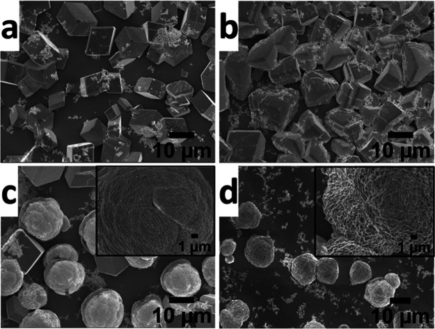
Figure 2.
Scanning electron microscopy images of CaCO3 crystals nucleated in the presence of PMMA-NPs at different concentrations of 25 μg/mL (a), 50 μg/mL (b), 75 μg/mL (c), and 125 μg/mL (d) under ambient conditions. The insets in (c) and (d) indicate the enlarged view of the single crystal surface. The SEM image of rhombohedral calcite crystals of CaCO3 obtained in the absence of PMMA-NPs is given in the Supporting Information (Figure S2).

