FIGURE 4.

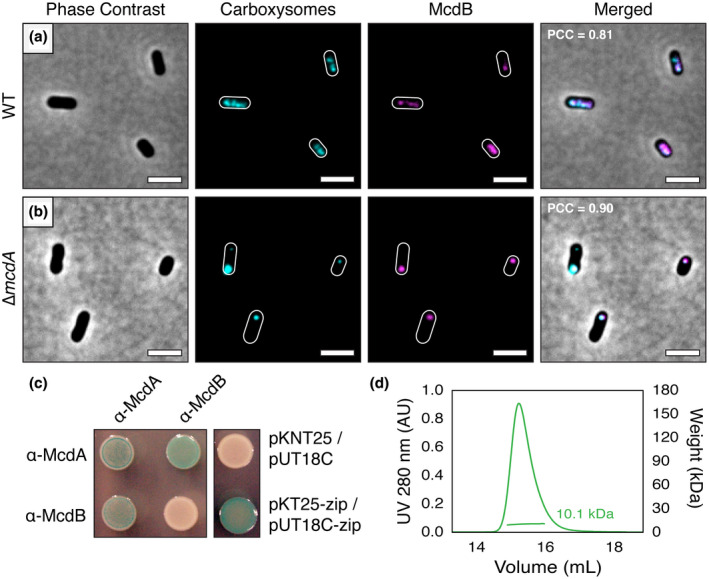
α‐McdB loads onto α‐carboxysomes and interacts with α‐McdA. (a) mNG‐McdB (magenta) colocalizes with the carboxysome reporter CbbS‐mTQ (cyan) in WT H. neapolitanus. (b) In the absence of McdA, McdB strongly colocalizes with carboxysome aggregates. PCC values were calculated from n ≥ 100 cells per cell population. Scale bar: 2 µm. (c) Bacterial‐2‐Hybrid (B2H) analysis of α‐McdA and α‐McdB. α‐McdA was positive for self‐association. α‐McdA directly interacts with α‐McdB. α‐McdB did not self‐associate. B2H image is representative of three independent trials. (d) SEC‐MALS plot for H. neapolitanus α‐McdB; monomer MW = 10 kDa [Colour figure can be viewed at wileyonlinelibrary.com]
